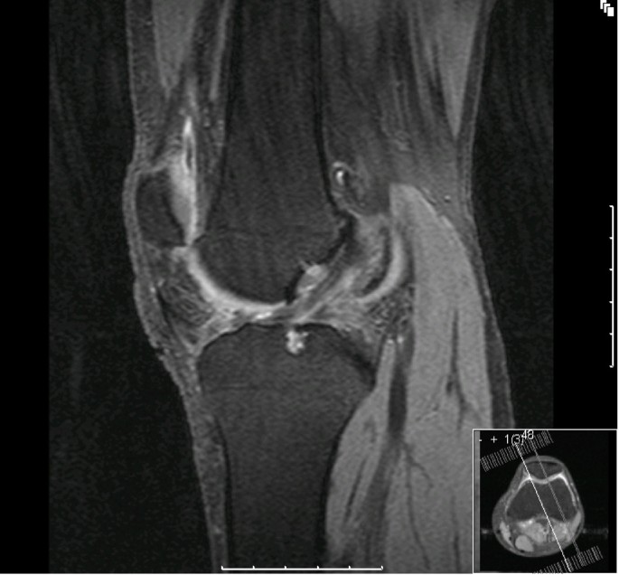
thin vertical peripheral tears of the meniscus or small corner tears of the meniscocapsular and meniscotibial ligaments with fluid interposition. non-compact tissue applied to the posterior horn of the medial meniscus that comprises the meniscocapsular ligament and the meniscotibial ligament with intervening fatty issue. To catch them, radiologists should become familiar with the normal appearance of the meniscocapsular junction of the posterior horn or the medial meniscus. These lesions are best seen on sagittal proton density-weighted and T2-weighted fat-saturated MR sequences. the peripheral part of the posterior horn or attachments to the posterior horn, particularly the meniscocapsular and meniscotibial ligaments.  vertical longitudinal tears located at the peripheral meniscocapsular junction of the posterior horn medial meniscus. There are five types of lesions, and they typically involve: This injury is most common with young male patients who play contact sports. In an article published June 17 in the American Journal of Roentgenology, a team from The Chinese University of Hong Kong outlined what these commonly missed injuries are and what providers should look for to detect them. Identifying them can also potentially help patient avoid further injury.
While catching these injuries might not change clinical management or lead to surgery, it can reduce patient symptoms, such as pain, tenderness, clicking, locking, and instability.

But, there are five knee injuries that are frequently overlooked, particularly by inexperienced readers. Knee injuries are common, and MRI is a highly accurate way to visualize the problem.


 0 kommentar(er)
